I started my Ph.D. four years ago when Trump became a president and finalized it during the pandemic. However, the adventure was beyond that...
Fetal and Neonatal Brain Segmentation


Important brain development in infants happens in the last trimester of pregnancy i.e., between 30 and 40 weeks of gestation. Monitoring brain developments during this period can help clinicians to detect abnormalities in the early stage. Magnetic Resonance Imaging (MRI) is a non-invasive tool that visualizes brain tissues. We can measure brain development from MRI in different stages or detect any abnormalities.
Brain Tissue Segmentation
A prerequisite to quantify brain development is to segment MRI into different brain tissue types. It's simply possible to manually go over every single slice of MRI and paint the brain into different tissue types, such as cortex, white matter, and gray matter. This job is categorized as one of the most boring jobs and it's extremely time-consuming. And here is where my research make sense, to develop an automatic algorithm to quantify brain development using machine learning and deep learning. The results of this research are published in AI special issue of MRI journal and ca be find here.

Tissue Segmentation in MRIs with Artifact


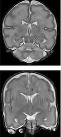

Recently, machine learning methods and particularly deep learning achieved excellent performance in many tasks. Many technologies from autonomous driving to face detection available in smartphone cameras are using these algorithms. Convolutional neural networks (CNNs) is the most popular network in image processing, classification, and segmentation tasks. This network shows high performance in medical image segmentation. However, the results are optimal in scans with artifacts (see the second column of the above figure). These scans visualize motion artifacts that are occurring due to infants' movement during the scanning. To overcome this challenge, an algorithm using another type of neural network recently widely used in image generation called generative adversarial network (GAN) is used. GAN composed of two networks, a generator, and a discriminator. We used this algorithm to remove motion artifacts from scans and improve brain tissue segmentation simultaneously. The results are published in MICCAI 2019 and can be find here.